THORACIC AORTA INCLUDING AORTIC
COARCTATION & DISSECTION
Pulse Sequences
1. BFFE Survey
2. Ref Scan
3. SS Axial TE 200
4. eTHRIVE Descending Aorta Axial BH
5. 3 D MRA pre Sagittal Plane
6. 3 D 512 MRA Sagittal Plane
7.
2D TIMING BOLUS CORONAL PLANE
8. 3 D 512 MRA Sagittal Plane
9. 3 D 512 MRA Sagittal Plane
10. e-THRIVE post Axial BH
11. FFE PROSET Axial Plane
12. SQFLOW_BH (Phase Contrast images of proximal and distal descending thoracic aorta as indicated below. For coarctation of the aorta, also obtain phase contrast image at level of the coarctation).
References & Illustrations
Ø
Standardized cardiovascular magnetic resonance
imaging (CMR) protocols, society for cardiovascular magnetic resonance: board
of trustees task force on standardized protocols http://scmr.org/assets/files/1532-429X-10-35.pdf
Ø Quantification of Flow Dynamics
in Congenital Heart Disease: Applications of Velocity-encoded Cine MR Imaging http://radiographics.rsna.org/content/22/4/895.full
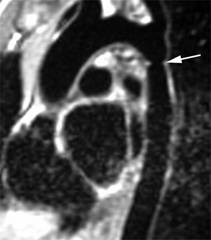
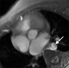
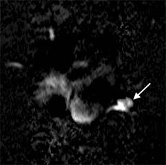
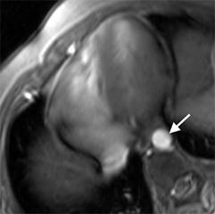
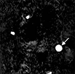
o Velocity-Encoded Cine MR Imaging For Quantification Of Collateral Circulation & Determining The Severity Of Aortic Coarctation- (Left) Oblique sagittal SE MR image show an aortic coarctation (top image). The bottom image shows how to obtain a velocity encoded image at the level of the coarctation (C) and the planes selected for velocity-encoded cine MR acquisitions for the proximal (P) and distal (D) thoracic aorta. Both planes are perpendicular to the direction of blood flow in the descending aorta. Middle images show magnitude and right images show phase images obtained from velocity-encoded acquisitions in proximal (top image) and distal (bottom image) locations of the descending aorta.

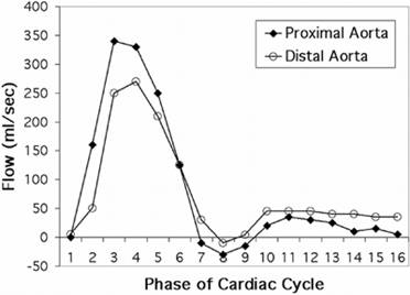
o Graph illustrates aortic flow in a patient without coarctation of the aorta. The flow curve is calculated by plotting blood flow in the proximal and distal aorta against time. In healthy individuals, flow in the proximal aorta is slightly greater than flow in the distal aorta.
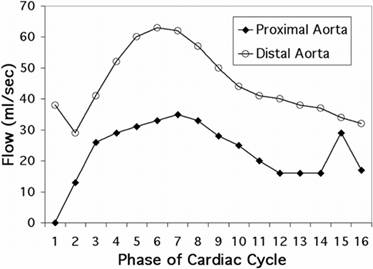
o Graph illustrates aortic flow in a patient with severe coarctation of the aorta. Patients with hemodynamically significant coarctation have greater flow distally than proximally due to collateral flow, which is quantified by subtracting proximal flow volume from distal flow volume. The patient in this case had twice as much blood flow in the distal aorta as in the proximal descending aorta, indicating that the volume of collateral circulation was approximately equal to flow just beyond the coarctation.
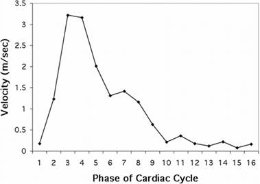
o Calculation of the pressure gradient across the coarctation. Graph illustrates how peak velocity across the coarctation is plotted against time. The pressure gradient across the coarctation is calculated with the modified Bernoulli equation ΔP= 4v2, where ΔP is the pressure gradient and v is the peak flow velocity. In this case, the peak velocity was 3.2 m/sec and the pressure gradient was 41 mm Hg, which is considered severe. The patient underwent surgical repair of the coarctation.
Phase Contrast Articles
Ø Cardiovascular Applications of Phase-Contrast MRI http://www.ajronline.org/cgi/content/full/192/3/662
Ø Cardiovascular Flow Measurement with Phase-Contrast MR Imaging: Basic Facts and Implementation http://radiographics.rsna.org/content/22/3/651.full
SVC and Inferior Vena Cava:
The encoding velocity for the first measurement or for
flow measurement is 110
cm/sec. The usual velocity for peak velocity measurement is 50–80 cm/sec.
Ascending Aorta:
The encoding velocity for the first measurement or
for flow measurement is 200 cm/sec. The usual velocity for peak velocity measurement is 100–160 cm/sec.
Main Pulmonary Artery:
The encoding velocity for the first measurement or
for flow measurement is 180 cm/sec. The usual velocity for peak velocity measurement is 60–120 cm/sec.
Right and Left Pulmonary Arteries:
The encoding velocity for the first measurement or
for flow measurement is 200 cm/sec. The usual velocity for peak velocity measurement is 60–120 cm/sec.
Ø Quantification of Flow Dynamics in
Congenital Heart Disease: Applications of Velocity-encoded Cine MR Imaging http://radiographics.rsna.org/content/22/4/895.full
Rev: 5/13/10