Post-Gadolinium Fat-Suppressed Single-Section Gradient Echo
After gadolinium administration, vessels
and enhancing tissues are shown best if signal from fat is suppressed. However,
in multi-section FMPSPGR images, 90-degree excitation
pulses targeted to nearby slices saturate flowing blood, reducing their signal
intensity. High signal intensity of vascular lumens is more uniform if only
one image section is obtained per acquisiton. To maintain efficiency, TR must
be reduced. The short TR also helps reduce sensitivity to motion, because
the center of k-space is sampled in less than a second. With current fat-saturated
techniques, the minimum TR for 2D gradient echo images is about 20 msec. To
prevent oversaturation of tissues, flip angle should be reduced to about 30
degrees.
Similar images can be obtained using 2D-TOF (time-of-flight),
but this technique uses flow compensation (gradient moment nulling), which
increases TE and therefore TR and acquisition time. With TE less than 3 msec.,
flow-induced signal loss is minimal, so flow compensation is not needed.
On some systems, fat saturation is not an option for
the single section pulse sequence database (FSPGR). FMPSPGR
with TR = 20 msec. results in single-section acquisition.
Images such as these are obtained as the final sequence
in nearly all body MRI examination, usually in the axial plane, showing delayed
extracellular (vascular + interstitial) space enhancement. Their bright blood,
dark fat and minimal motion artifact results in a resemblance to contrast-enhanced
CT images.
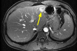 |
Post-gadolinium fat-suppressed single-section gradient echo image with TR/TE/flip = 20/2.1/30. All vessels have high signal intensity, due to a combination of gadolinium-enhancement and time-of-flight effects. Arrow indicates cavernous hemangioma. |
Copyright © Thomas Jefferson University. All Rights Reserved.
The Thomas Jefferson University web site, its contents and programs, is provided for informational and educational purposes only and is not intended as medical advice nor is it intended to create any physician-patient relationship. Please remember that this information should not substitute for a visit or a consultation with a health care provider. The views or opinions expressed in the resources provided do not necessarily reflect those of Thomas Jefferson University, Thomas Jefferson University Hospital, or the Jefferson Health System or staff.