Benign Adrenal Adenoma
Adrenal Metastasis

|
Benign Adrenal Adenoma
|
Adrenal Metastasis
|
||
|
|
|
|
|
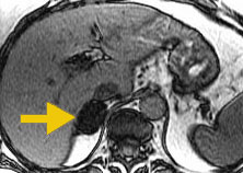
In this T1-weighted image, fat and water signals are additive. Arrow = adrenal adenoma. |
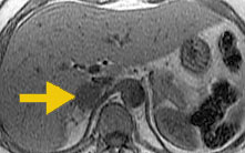
In this T1-weighted "opposed phase" image, fat and water signals interfere with each other. The adrenal adenoma (arrow) contains fat and water, so its signal intensity has decreased. A metastasis is therefore excluded. |
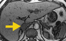
In this T1-weighted image, where fat and water signals
are additive, the adrenal metastasis (arrow) is slightly less intense
than in the adenoma above.
|
In this T1-weighted "opposed phase" imager, the adrenal metastasis (arrow) has the same signal intensity as in the in-phase image to the left. This is because the metastasis does not contain fat. |
1. Mitchell DG, Crovello M, Matteucci T, R.O. P, Miettinen MM. Benign adrenocortical masses: diagnosis with chemical shift MR imaging. Radiology 1992;185:345-351.
2. Outwater EK, Siegelman ES, Radecki PD, Piccoli CP, Mitchell DG. Distinction between benign and malignant adrenal masses: value of T1-weighted chemical shift MR imaging. AJR 1995;165:579-583.
 |
Jump to Department
of Radiology Home Page ( Leave MRI site) |