|
|
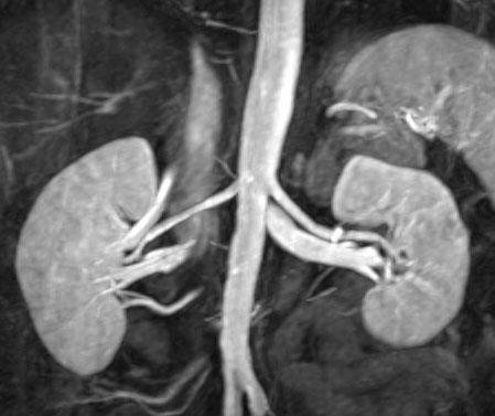
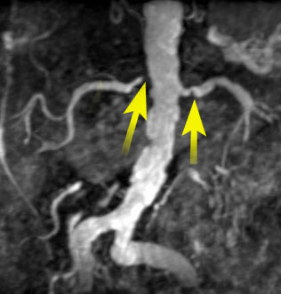
| In this 3D coronal gadolinium-enhanced MRA, the kidneys and renal arteries appear normal, and there is no aortic aneurysm. | In a patient with severe atherosclerosis, arrows indicate bilateral renal artery stenosis |
1. DeCobelli F, Vanzulli A, Sironi S, et al. Renal artery stenosis: Evaluation with breath-hold, three-dimensional, dynamic, gadolinium-enhanced versus three-dimensional, phase-contrast MR angiography. Radiology 1997;205:689-695.
2. Prince MR, Schoenberg SO, Ward JS, Londy FJ, et al. Hemodynamically significant atherosclerotic renal artery stenosis: MR angiographic features. Radiology 1997;205:128-136.
3. Prince MR. Peripheral vascular MR angiography: the time has come. Radiology 1998;206:592-593.
4. Quinn SF, Sheley RC, Semonsen KG, Leonardo VJ, et al. Aortic and lower-extremity arterial disease: evaluation with MR angiography versus conventional angiography. Radiology 1998;206:693-701.
 |
Jump to Department
of Radiology Home Page ( Leave MRI site) |