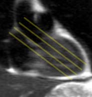
Coronal subsecond T2-weighted single-shot fast spin
echo localizer, used to prescribe the acquisition of ventricular long
axis images.
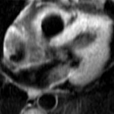
Long axis blood-suppressed image at the level of the
right ventricular outflow tract, acquired during suspended respiration.
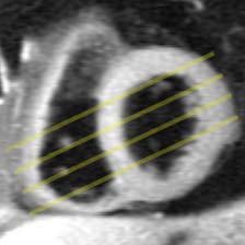
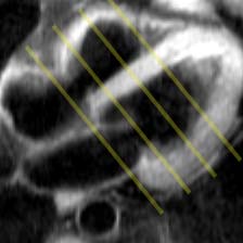
Long axis blood-suppressed image at the level of the
mitral and tricuspic valves, used to presecribe the short axis acquisition.
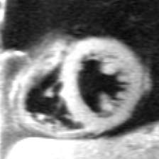
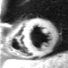
Short axis blood-suppressed image at the distal ventricles.