|
|
|
|
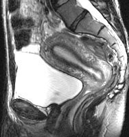
In this sagittal T2-weighted image, the endometrium is
depicted clearly because its water content is higher than that of the
myometrium, especially the inner myometrium.
|
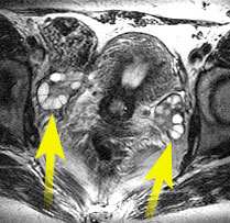
In this transverseT2-weighted image, the ovaries (arrows)
are depicted clearly because of the fluid within the follicles.
|
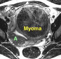
In this T2-weighted image of a different patient, a myoma
is depicted as low signal intensity with well-defined margins. In contrast,
an area of focal adenomyosis (A) is less well-defined, blending with the
inner myometrium.
|
More than any other method of imaging, MRI allows identification of normal anatomy and a diversity of abnormalities, including myomas, adenomyosis, endometriosis and malignancy.
1. Outwater EK, Siegelman ES, Wilson KM, Mitchell
DG. Benign and malignant gynecologic disease: Clinical importance of fluid
and peritoneal enhancement in the pelvis at MR imaging. Radiology 1996;200:483-488.
2. Outwater EK, Siegelman ES, Van Deerlin V. Adenomyosis: current concepts and imaging considerations. AJR 1998;170:437-441.
 |
Jump to Department
of Radiology Home Page ( Leave MRI site) |