|
|
|
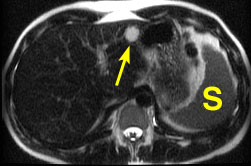
Heavily T2-weighted image shows a well defined liver lesion
(arrow) that is much brighter than the spleen (S). A malignant
lesion would have signal intensity similar to or less than that of
the spleen.
|
|
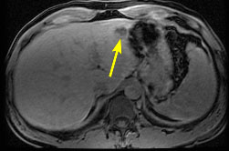
Unenhanced T1-weighted fat suppressed image.
|
|
|
|||
 |
|
||
|
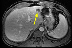
T1-weighted fat suppressed image about 30 seconds after
intravenous injection of gadolinium contrast agent. There is some nodular
enhancement at the periphery of the lesion.
|
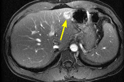
About 5 minutes after injection of gadolinium contrast
agent, most of the lesion has enhanced. Progressive nodular enhancement
is only seen with cavernous hemangiomas, not malignant masses.
|
References
1. Ito K, Mitchell DG, Outwater EK, Szklaruk J, Sadek AG. Hepatic lesions: Discrimination of nonsolid, benign lesions from solid, malignant lesions with heavily T2-weighted fast spin-echo MR imaging. Radiology 1997;204:729-737.
2. Semelka RC, Worawattanakul S, Kelekis NL, et al. Liver lesion detection, characterization, and effect on patient management: comparison of single-phase spiral CT and current MR techniques. JMRI 1997;7:1040-1047.
 |
Jump to Department
of Radiology Home Page ( Leave MRI site) |