|
|
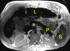
| The Liver (L) and pancreas (P) have decreased signal intensity due to increased iron. The spleen (S) is normal. This is the pattern typical of idiopathic hemochromatosis. |
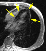
In the same patient, myocardial iron is increased (arrows). |
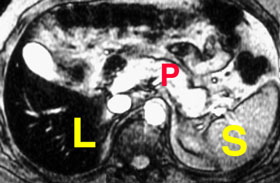
In a different patient, with transfusional siderosis, iron is increased in the liver (L) and spleen (S), the principal reticuloendothelial organs. The pancreas (P) is normal. (Previously published in reference #3) |
 |

|
|
In this 35 year old asymptomatic man with a family history
of idiopathic hemochromatosis, MRI shows abnormally low signal intensity
isolated to the liver (L). The spleen is normal, typical of hemochromatosis.
The pancreas is not yet affected at this early stage of disease. (Previously
published in reference #1)
|
One year later, after monthly phlebotomy, MRI shows nearly
normal liver signal intensity, confirming successful reduction of body
iron stores. (Previously published in reference #1)
|
1. Siegleman ES, Mitchell DG, Semelka RC. Abdominal iron deposition: metabolism, MR findings, and clinical importance. Radiology 1996;199:13-22.
2. Siegelman ES, Mitchell DG, Outwater E, Munoz SJ, Rubin R. Idiopathic hemochromatosis: spectrum of MR findings in cirrhotic and precirrhotic patients. Radiology 1993;188:637-641.
3. Mitchell DG. Case 16 - Idiopathic Hemochromatosis. In Mirowitz SA, Kneeland JB, Siegel MJ, Tempany CMC, Weinreb JC, White CS: ACR Body MRI Test and Syllabus; 46:201-212.
 |
Jump to Department
of Radiology Home Page ( Leave MRI site) |