|
Most reports indicate
that MRI is more sensitive and more specific than CT or other tests for
detecting liver malignancies and for distinguishing them from benign lesions.
|
|
|
|
|
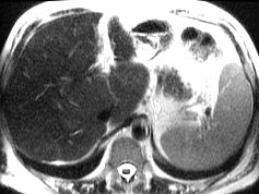
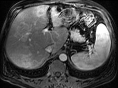
T2-weighted image does not show any liver lesions. Benign
lesions such as cavernous
hemangioma are much brighter.
|
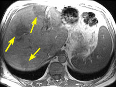
In this T1-weighted, three liver lesions are identified at this level (arrows). |
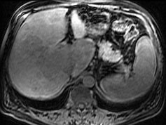
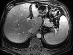
Fat signal has been suppressed in this T1-weighted image.
The liver has lost some signal because of mild fatty infiltration, so
the liver lesions are not seen as well.
|
|
|
||
 |
 |
|
|
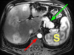
With MRI technique similar to that used above right, but
immediately after rapid intravenous injection of gadolinium contrast agent,
liver metastases enhance intensely. Intense enhancement is also seen in
aorta (red arrow), pancreas (green arrow) and spleen (S).
|
About 30 seconds after gadolinium contrast agent injection, liver lesions are subtle. |
About one minute after gadolinium contrast agent injection,
liver lesions are no longer visible.
|
1. Ito K, Mitchell DG, Outwater EK, Szklaruk J, Sadek AG. Hepatic lesions: Discrimination of nonsolid, benign lesions from solid, malignant lesions with heavily T2-weighted fast spin-echo MR imaging. Radiology 1997;204:729-737.
2. Semelka RC, Worawattanakul S, Kelekis NL, et al. Liver lesion detection, characterization, and effect on patient management: comparison of single-phase spiral CT and current MR techniques. JMRI 1997;7:1040-1047.
 |
Jump to Department
of Radiology Home Page ( Leave MRI site) |