In-situ mucin-hypersecreting intraductal adenocarcinoma (low malignant potential)
|
|
|
|
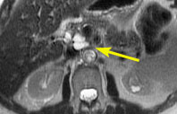
In this sub-second transverse T2-weighted image, there
is a complex cystic mass in the head of the pancreas (arrow).
|
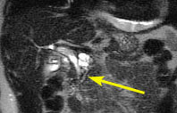
In this sub-second coronal T2-weighted image, the the
cystic mass is shown to communicate with the pancreatic duct (arrow),
which is mildly dilated.
|
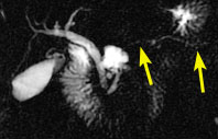
Sub-second coronal-oblique MRCP image shows the normal
biliary system, duodenum, gallbladder, and the cystic mass communicating
with the pancreatic duct. The duct in the pancreatic tail (small arrows)
is not dilated.
|
1. Fulcher AS, Turner MA. Magnetic resonance pancreatography: experience in 400 patients. Radiographics 1998.
2. Holzknecht N, Gauger J, Sackmann M, Thoeni RF, et al. Breath-hold MR cholangiography with snapshot techniques: prospective comparison with endoscopic retrograde cholangiography. Radiology 1998;206:657-664.
3. Chan Y, Chan ACW, Lam WWM, Lee DWH, et al. Choledocholithiasis: Comparison of MR cholangiography nd endoscopic retrograde cholangiography. Radiology 1996;200:85-89.
 |
Jump to Department
of Radiology Home Page ( Leave MRI site) |