|
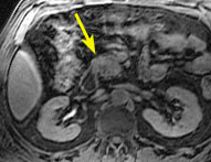
Small Adenocarcinoma
Without Vascular Invasion
|
||
![]() |
![]() |
![]() |
|
In this sub-second coronal T2-weighted image, there is
a solid mass in the head of the pancreas (arrow).
|
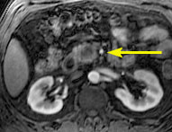
In this sub-second coronal MR cholangio-pancreatography
image, the the mass is shown to obstruct the pancreatic duct (arrow).
|
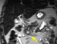
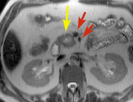
Sub-second transverse T2-weighted image shows the mass
(yellow arrow), which does not involve the superior mesenteric artery
and vein (red arrows).
|
![]() |
![]() |
![]() |
|
Ttransverse fat suppressed T1-weighted image shows the
mass (arrow).
|
Immediately after injection of gadolinium contrast agent,
the mass shows reduced enhancement. Arrow incidates the uninvolved superior
mesenteric artery.
|
Approximately 30 seconds after injection of contrast agent,
the superior mesenteric vein is shown (arrow).
|
|
More than any other method
of imaging, MRI can depict pancreatic masses and their relationship to
biliary and pancreatic ducts, peripanreatic vessels and surrounding tissues.
|
||
References
1. Mammone JF, Siegelman ES, Outwater EK. Magnetic resonance imaging of the pancreas and biliary tree. Seminars in Ultrasound, CT and MR 1998;19:35-52.
2. Ichikawa T, Haradome H, Hachiya J, Nitatori T, et al. Pancreatic ductal adenocarcinoma: preoperative assessment with helical CT versus dynamic MR imaging. Radiology 1997;202:655-662.
3. Semelka RC, Kelekis NL, Polina PL, Sharp T, Calvo B. Pancreatic cancer detection: comparison of spiral CT and MR imaging by using ROC analysis. Radiology 1995;197(P):378.
4. Mitchell GG, Winston CB, Outwater EK, Ehrlich SM. Delineation of pancreas with MR imaging: multiobserver comparison of five pulse sequences. JMRI 1995;5:193-199.
5. Mitchell DG. MR imaging of the pancreas. MRI Clinics of North America 1995;3:51-71.
![]() |
Jump to Department
of Radiology Home Page ( Leave MRI site) |