|
|
|
|
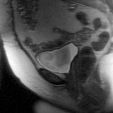
Sagittal T2-weighted image. The bladder has high signal
intensity.
|
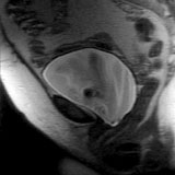
Sagittal T2-weighted image obtained during straining.
Note descent of the bladder.
|
The patient was instructed to alternately strain and relax
as a series of sub-second T1-weighted images were obtained, showing the
dynamic motion of the pelvic structures with straining.
|
 |
Jump to Department
of Radiology Home Page ( Leave MRI site) |