Arrhythomgenic Right Ventricular Dysplasia

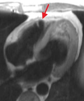
Long axis blood suppressed image obtained at diastole during suspended respiration. The arrow indicates thinning of the right ventricular apical wall.
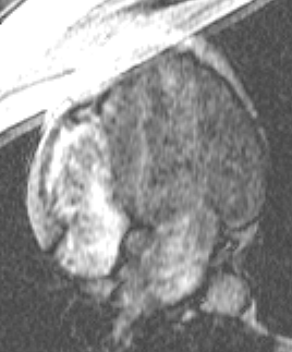
Long axis cine image obtained during suspended respiration shows dyskinesia of the right ventricular apex.
 
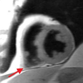
Short axis blood suppressed image obtained at diastole during suspended respiration. The arrow indicates thinning of the right ventricular apical wall.
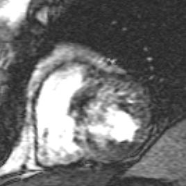
Short axis cine image obtained during suspended respiration shows dyskinesia of the right ventricular apex.
MRI is the best method for evaluating right ventricular wall morphology and function.

Jump to Department of Radiology Home Page ( Leave MRI site)
Copyright © Thomas Jefferson University. All Rights Reserved.
The Thomas Jefferson University web site, its contents and programs, is provided for informational and educational purposes only and is not intended as medical advice nor is it intended to create any physician-patient relationship. Please remember that this information should not substitute for a visit or a consultation with a health care provider. The views or opinions expressed in the resources provided do not necessarily reflect those of Thomas Jefferson University, Thomas Jefferson University Hospital, or the Jefferson Health System or staff.