MR CholangioPancreatography (MRCP)
A heavily T2-weighted technique that emphasizes
signals from simple fluid, such as bile and pancreatic juice. The SSFSE
localizer sequence that begins most abdominal MRI exams can serve as a tomographic
MRCP sequence. The moderate TEef of about 180 msec. and the lack of fat suppression
on these images allows signal from background tissue to provide anatomic landmarks.
If maximum intensity projection (MIP) images are to be obtained, higher TEef
and fat suppression should be used.
Using an axial image, multiple coronal oblique images are
obtained radially oriented around the pancreatic head. Additional oblique
images should be obtained oriented along the pancreatic body/tail. With TEef
nearly 1000 msec, only signals from fluid are depicted. Image thickness should
be about 4 - 5 cm., and field of view should be about 25 cm. This technique
provides high-resolution MRCP images with minimal motion sensitivity and no
need for MIP reconstructions.
Each image is obtained in less than one second. However,
at least 10 seconds should elapse between each image, to prevent severe degradation
caused by cross-talk from the overlapping thick sections.
 |
 |
 |
|---|---|---|
|
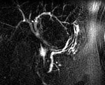
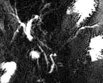
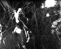
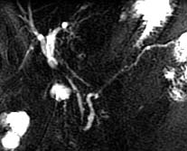
Axial SSFSE image above is
used to prescribe thick overlapping sections, oriented radially around
the pancreatic head (yellow), and the pancreatic tail (green). Four
of the MRCP images are shown at right.
|
 |
 |